


Circulating Tumor Cells (CTCs)
CTCs are a subpopulation of aggressive tumor cells that can migrate from the primary tumor through the blood vessels to reach distal organs and form metastases.
CTCs are able to strongly modify their phenotype to survive in the blood vessels. They become resistant to anoikis, the programmed cell death that occurs when a cell detaches from a solid structure, evade immune surveillance, extravasate into a distal organ, and ultimately to form metastases.
Not much is known about these cells, particularly because they are very rare and very difficult to isolate from patients’ blood samples. Understanding the mechanisms behind their formation and survival in the bloodstream, would greatly advance our knowledge of metastases biology and could aid in the development of a new generation of drugs capable of blocking metastasis formation, thereby significantly reducing cancer mortality.
Furthermore, CTCs could become a valuable source for liquid biopsies, especially in cancers where surgery is not feasible.
After preliminary in vitro investigations, in which we studied the effects of hypoxia and radiation on the primary tumor in generating a subpopulation of cells with CTCs-like phenotype, our group began exploring new methods to isolate CTCs from in vivo models.
Successfully isolating these cells has allowed us to collect new data on their phenotype and transcriptomic profile. Initial results indicate that CTCs are highly heterogeneous, but they all show significant differences in gene expression compared to the primary tumor (figure 2).
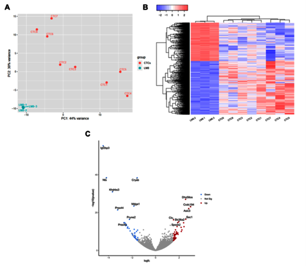
Figure 2: Gene expression comparison between parental and CTC-derived cell lines. (A) PCA plot showing all CTC-derived cell lines and three replicates of LM8, CTCs of 2 different experiments were analyzed. (B) Heatmap showing all DEGs (p-adj < 0.05) between the parental LM8 cell line and all CTC-derived cell lines, depicted are log2 (value + 1) transformed z-scores, complete Euclidean clustering. (C) Volcano plot showing DEGs between the parental cell line LM8 and CTC-derived cell lines, colors indicate a log(Fc) higher than 1.5 and a p-adj. < 0.05, 15 most differentially expressed Genes were labeled. Benje, M.; Vitacchio, T.; Fritsche, D.; Tinganelli, W. Gene Expression Profiling and Phenotypic Characterization of Circulating Tumor Cells Derived from a Murine Osteosarcoma Model. Cancers 2025, 17, 1210. https://doi.org/10.3390/ cancers17071210
Our next step is to identify the key genes responsible for CTC generation using gene knockout technologies.
Additionally, we will study the effects of radiation (comparing low versus high LET radiation, fractionation versus hypofractionation schemes, UHDR versus conventional dose rate irradiation) on the generation of CTCs.