


AI methods in particle therapy
It is ideal to have imaging, planning and treatment in a single session. The treatment planning process consists of multiple steps, and the patient must receive the treatment as soon as possible to prevent anatomical changes that may occur after the planning begins and can cause errors during the treatment delivery. Some steps, such as contouring, can be significantly accelerated by artificial intelligence. In contrast, others still require substantial computational resources, utilising the gold standard Monte Carlo simulation or an analytical algorithm as an approximation.
In the group, we integrate AI into various steps of our in-house analytical pencil beam algorithm, TRiP98, which aims to speed up or improve the results of the analytical calculation.
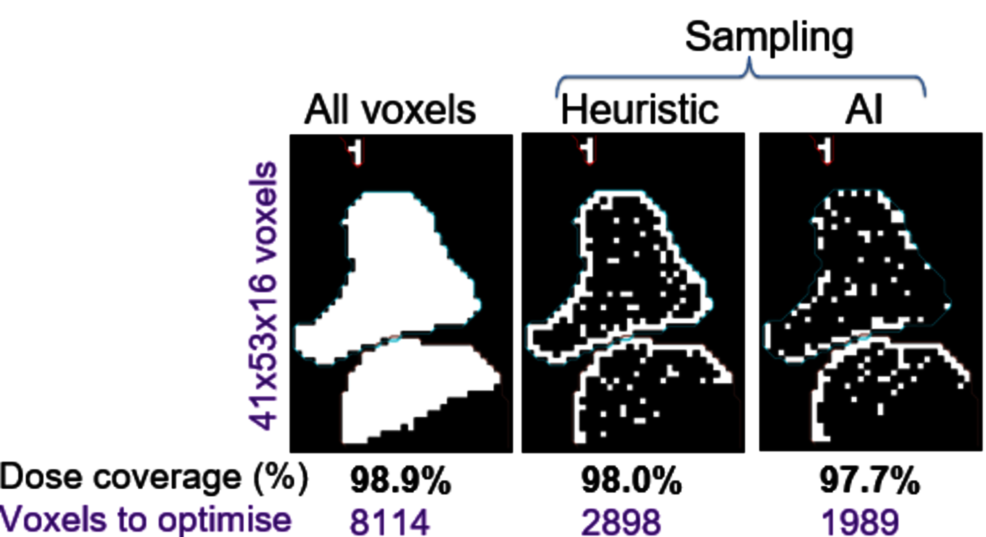
Voxel optimisation
Depending on the tumour size and treatment parameters, the number of optimisation points can require a significant amount of computational resources to optimise the plan and calculate the dose distribution related to it. Subsampling of voxels allows to speed up the calculation because of the reduction of the optimisation points. But the results of such optimisation can aggravate the quality of the optimisation. The voxels involved in optimisation should be chosen based on their location to provide a fast, yet quality-loss-free, optimised plan. We apply a deep learning model to select the most relevant voxels for the optimisation.
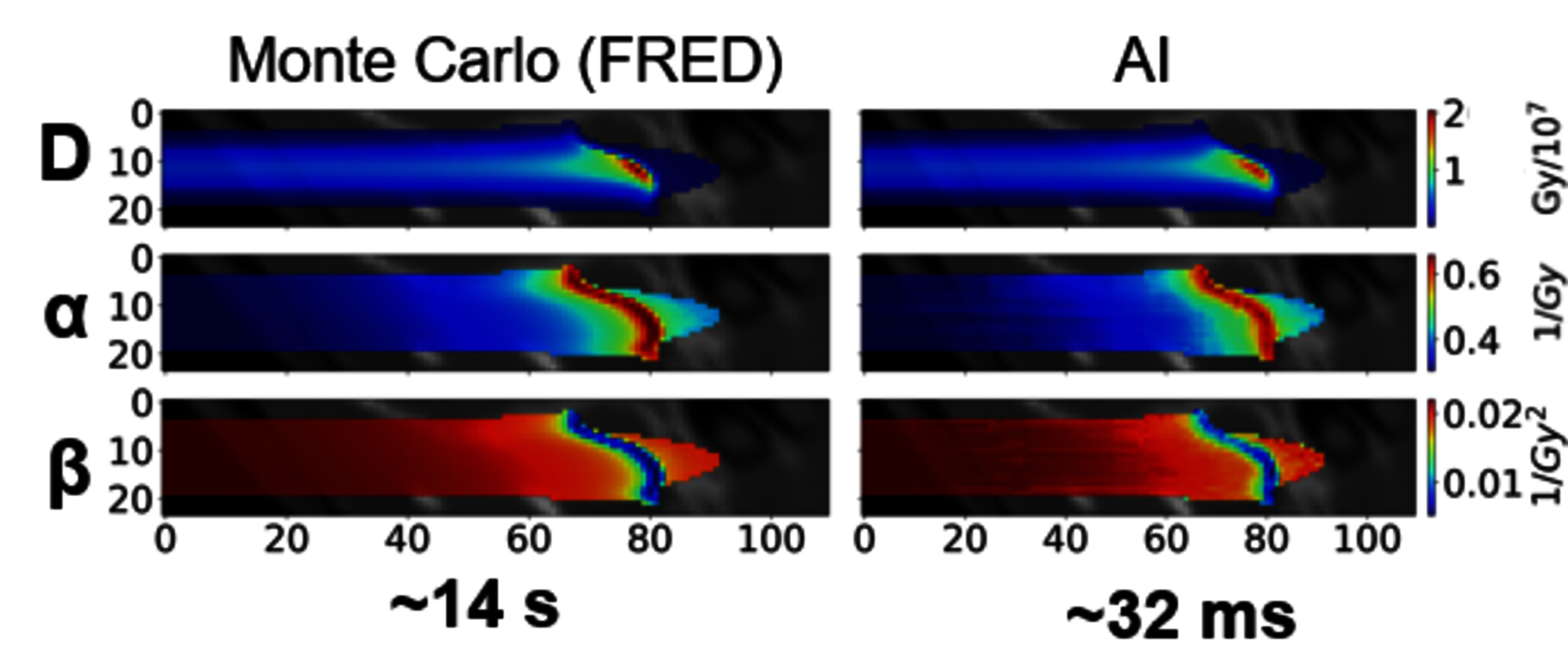
Dose calculation
A simulation of the heavy ions in a patient CT is a complex and time-consuming process, which does not permit the application of Monte Carlo methods in clinical routine. Instead, an analytical pencil beam algorithm is used. It delivers a good quality, fast approximation of the parameters required for biological effectiveness (RBE)–weighted optimisation and dose calculation in carbon ion therapy , but struggles with inhomogeneous tissues. To overcome the drawback but keep the benefit of its speed, we developed a transformer-based model to compute the parameters with MC quality in milliseconds.
Mixed beam range prediction
The PROMISE project employs a mixed helium-carbon beam for concurrent treatment and real-time imaging. The scintillator detector captures both the helium Bragg peak and carbon fragmentation background, which interferes with conventional ion imaging algorithms for water equivalent thickness reconstruction. We apply deep learning methods to accurately suppress this background noise.
Additionally, we investigate algorithms to establish patient-specific helium-carbon range correlations. By measuring the helium Bragg peak, we can infer breathing phase and carbon dose delivery, enabling online range and dose monitoring. This approach allows for adaptive treatment planning when necessary.