


New results from the ERC project BARB on image-guided tumor therapy
28.07.2026 |
Using radioactive ion beams and in‑beam PET, researchers at GSI/FAIR, in collaboration with the Ludwig Maximillian Universität (LMU) Munich, are working on a new generation of highly precise, adaptive particle therapy. Because tumors and surrounding organs can shift or change shape during the course of treatment, radiotherapy plans often need to be adapted in real time to keep delivering the dose safely and effectively to the tumor. A new study within the ERC project „BARB – Biomedical Applications of Radioactive ion Beams“ shows that the range of a radioactive ¹¹C ion beam in a mouse model can be deliberately varied and monitored in real time – from short, right to long range settings. This work provides potential of radioactive ion beams for real-time adaptive radiotherapy, and shows that the beam range selected by online imaging directly affects the biological outcome.
Marco Durante, head of the Biophysics Department and PI of the ERC AdG BARB noted that “BARB has transformed radioactive ion therapy from a visionary idea, already explored in the past in different centers including fragment separator FRS of GSI, into an experimentally validated technology. By establishing the scientific principles, demonstrating feasibility under realistic conditions and identifying practical routes towards implementation, the project has laid the foundations for a new generation of image-guided particle therapy and has reinforced GSI/FAIR leading position in biomedical applications of accelerator science.”
Christoph Scheidenberger, head of the FRS group, said: “We are happy that our radioactive ions and detectors enable us to make a significant contribution to this successful medical research. The FRS was developed to produce and study new isotopes in order to understand element formation in stars and was recently used to produce the carbon-11 isotopes required for medical studies. This demonstrates the importance of interdisciplinary work in engineering and science, as practiced at large research facilities such as GSI and, in the future, at FAIR—and on an even larger scale at the new Super-FRS facility of FAIR.“
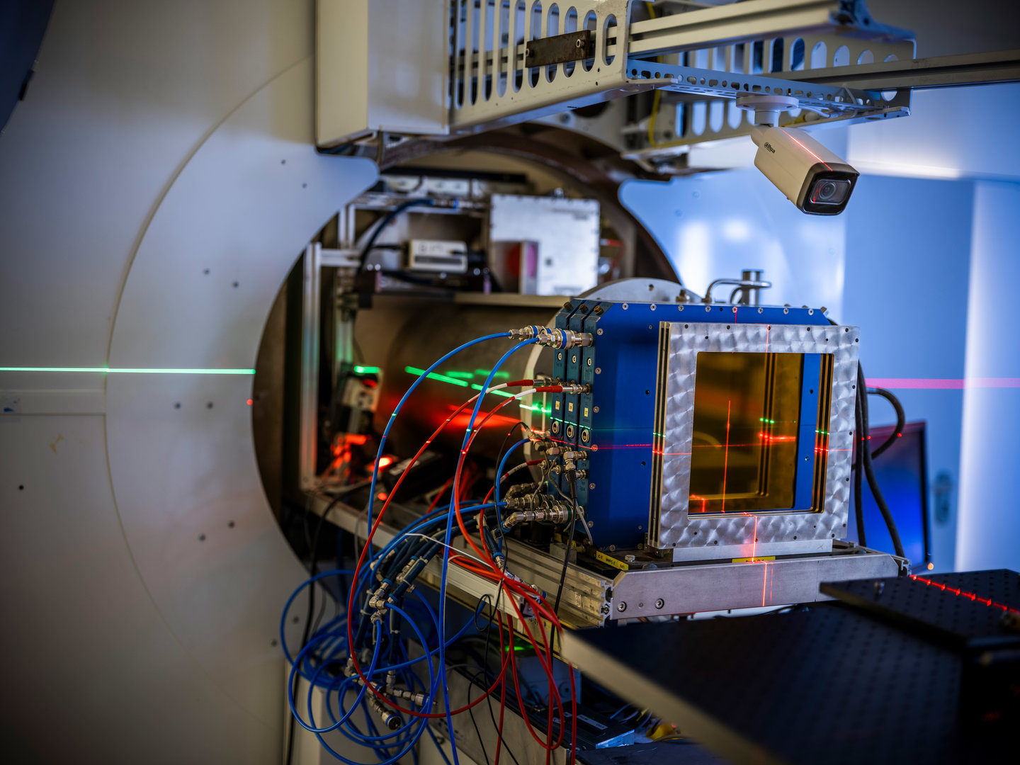
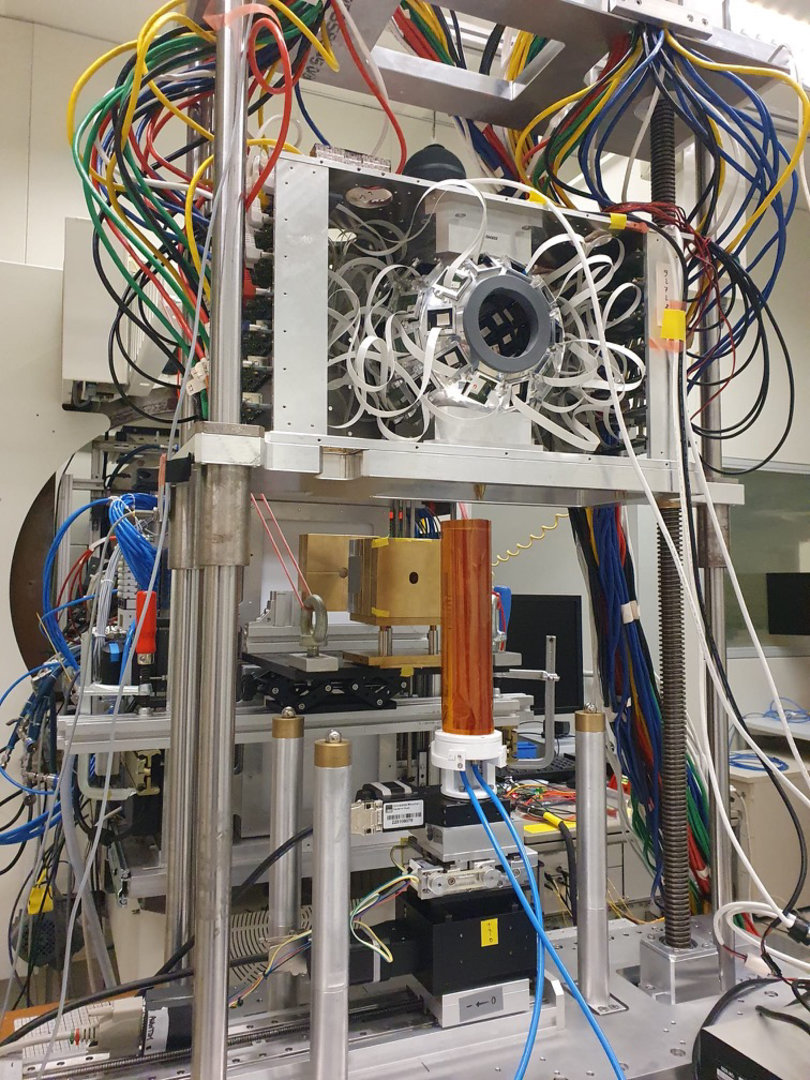
The focus of the study published in Communications Medicine is whether radioactive ion beams can serve as a tool for adaptive particle therapy. To this end, a mouse model with a neck tumour was irradiated: a narrow, quasi‑monoenergetic ¹¹C probing beam was first used to determine the beam range with sub‑millimetric precision using in‑beam PET. A therapeutic spread‑out Bragg peak distribution was then delivered at the same position. Three beam depths were tested: a too‑short beam that stops at the front edge of the tumour, an optimal “Goldilocks” range that completely covers the tumour while sparing healthy tissue behind it, and a too‑long beam that traverses the tumour, spinal cord and esophagus. The results are clear: only in the optimal “Goldilocks” range was complete tumour control achieved without measurable damage to spinal cord or esophagus; with a too‑short range residual tumour remained, and with a too‑long range marked damage to the esophagus and nervous system occurred.
Background: first image‑guided treatment with ¹¹C in Nature Physics
The new results build on an earlier study published in Nature Physics in 2025. In that work, the BARB team showed for the first time that a neck tumour can be successfully treated with a radioactive ¹¹C ion beam, even though the tumour is located very close to the sensitive spinal cord. The beam served simultaneously for therapy and as a tracer for a high‑resolution in‑beam PET scanner, which allowed real‑time monitoring of the beam range during irradiation. This experiment provided the first evidence that image‑guided therapy with radioactive ion beams is feasible, safe and effective under realistic conditions.
Radioactive ions from the Fragment Separator FRS
Both studies were enabled by the Fragment Separator FRS at the GSI/FAIR accelerator and experiment complex. There, short‑lived radioactive ion beams are produced from a primary heavy‑ion beam and separated according to mass and charge, so that an intense ¹¹C beam can be provided specifically for biomedical experiments. The work carried out in BARB is therefore a clear example of close interdisciplinary collaboration between several GSI/FAIR research groups – from nuclear physics and beam delivery through medical physics to radiation biology – all pursuing the common goal of making particle therapy of the future even more precise and safe. GSI/FAIR also engage in collaboration with external research groups, and in fact BARB was made possible by the collaboration with LMU that built the PET for small animals used in the experiments. (IP)
More info
Paper in Communications Medicine


