The target online-diagnosis at SHIP
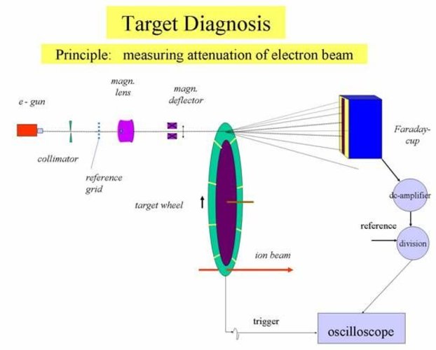
For the experiments at the SHIP-apparatus 8 sectors of banana shaped targets are mounted on a wheel rotating with a velocity of ~20 m/s and ~ 19 rps (figure 1). Target changes and damages due to irradiation by heavy ions are controlled using an electron beam from an electron gun. The electron beam penetrates the targets at the opposite of the ion beam path and is received by a slit shaped Faraday cup (figure 1). The electron signal is attenuated due to angular scattering and absorption of electrons in the material corresponding to the thickness of the target. Current fluctuations of the gun are compensated by normalizing the Faraday cup signal with the signal from a reference grid passed by the electrons in front of the target. A focusing magnetic lens defines the resolution of presently ~ 0.3 mm at the target. The target area is scanned in radial direction with a precision of < 0.1 mm by using a magnetic deflector. Scans along the circumference of the target wheel with a position resolution of < 0.1 mm results from the rotation where a trigger pulse starts the time base of an oscilloscope. The display of the oscilloscope is stored in a personal computer for handling the measured spectra. Thereby individual targets and areas at the rotating wheel can be displayed by defined delays switched to the trigger pulse. This set up allows a fast inspection of all targets within 60 ms without disturbing a running experiment.
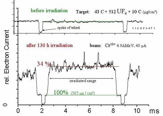
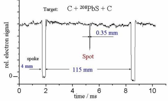
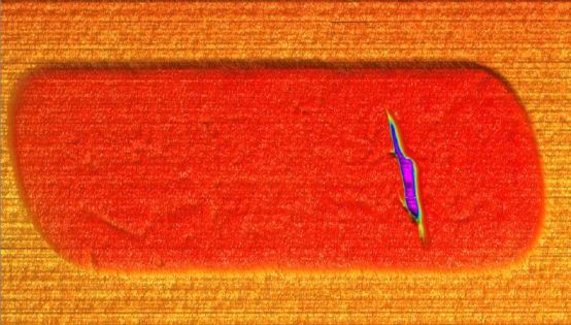
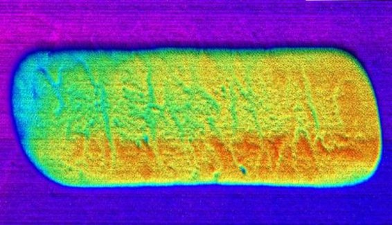
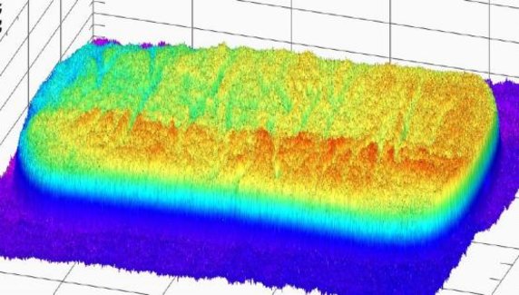
The method allows not only a permanent watching of changes induced by ion beam impacts but also contaminations, pin holes and variation of thickness from the production process itself can be detected. This is of relevance for nuclear reactions products which are favourably produced in a small energy window requiring a homogeneous energy loss in the target in order to receive the best efficiency through the SHIP filter. Also background reaction products can be strongly produced by impurities on the target (grains, dust particles,...) which can be made visible by the electron beam diagnosis.